Microscope
ProductOverview
The compound microscope magnifies small specimens up to about 1000× by passing light through the sample and a series of lenses. This trinocular laboratory model adds a third optical path to the binocular eyepieces, feeding a digital camera so the live image can be captured or displayed. It is a workhorse for histology, microbiology, and materials inspection. Köhler illumination, the defining feature of a research-grade stand, produces an evenly lit field free of any image of the lamp filament.
Construction
Everything mounts on the Stand & Frame, a heavy die-cast base and rigid C-shaped arm that gives the instrument a low center of gravity and holds the optical groups in fixed alignment. The specimen sits on the Mechanical Stage, a mechanical XY platform whose coaxial knobs translate the slide precisely under the objective through a rack and pinion. The Focus Mechanism mechanism raises and lowers the stage on a dovetail slide with stacked coarse and fine knobs, the fine knob geared down for micron-level adjustment.
How it works
The Illumination System system sets up the Köhler path: an LED lamp feeds a collector lens and field diaphragm, and the Condenser Assembly cones that light onto the slide with an adjustable aperture that controls contrast, resolution, and depth of field. Light passing through the specimen enters whichever objective is clicked into place on the Revolving Nosepiece, a detented turret carrying parfocal objectives from 4× to 100×, each correcting aberrations for a sharp magnified image.
The magnified beam travels up to the Trinocular Head, where a prism set splits it between the two eyepieces and the third camera tube. Through the Camera Port port, a C-mount adapter projects that third path onto a digital image sensor, and the optional Digital Display shows the live view on an LCD. A switch-mode power supply drives the lamp, camera, and display from mains.
Microscope parts and their functions
10 top-level parts · 60 parts in total · full bill of materials below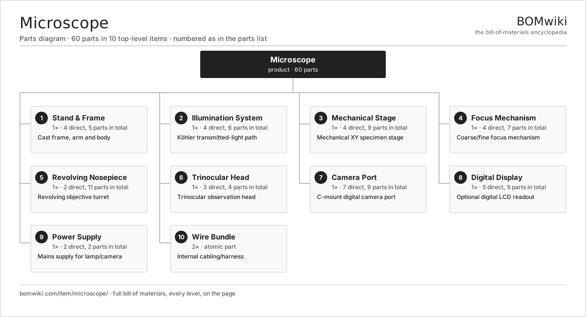
| # | Part | Qty | What it does |
|---|---|---|---|
| 1 | Stand & Frame 4 parts | 1× | Cast frame, arm and body |
| 2 | Illumination System 4 parts | 1× | Köhler transmitted-light path |
| 3 | Mechanical Stage 4 parts | 1× | Mechanical XY specimen stage |
| 4 | Focus Mechanism 4 parts | 1× | Coarse/fine focus mechanism |
| 5 | Revolving Nosepiece 2 parts | 1× | Revolving objective turret |
| 6 | Trinocular Head 3 parts | 1× | Trinocular observation head |
| 7 | Camera Port 7 parts | 1× | C-mount digital camera port |
| 8 | Digital Display 5 parts | 1× | Optional digital LCD readout |
| 9 | Power Supply 2 parts | 1× | Mains supply for lamp/camera |
| 10 | Wire Bundle | 2× | Internal cabling/harness |
Diagrams & photos

3D model
No 3D model yet. Have a CAD file of this? Add a 3D model. STL renders right on this page; STEP, FreeCAD, and OpenSCAD sources are welcome too (openly licensed, like all 3D models on BOMwiki).
Build & assembly graph
expand / collapse · shared sub-assemblies converge · links to related products · est. labourTap an assembly to expand/collapse · tap a part to open it · use “Open page” for any node · drag to pan, scroll to zoom.
Bill of materials for Microscope
10 top-level lines · 60 rows shown · 60 parts total · indented to 3 levels| # | Item / sub-assembly | Part no. | Qty/assy | Ext. qty | Parts | Type |
|---|---|---|---|---|---|---|
| 1 | Stand & Frame 4 parts | microscope-stand | 1× | 1 | 5 | assembly |
| 1.1 | Cast Base | microscope-cast-base | 1× | 1 | · | part |
| 1.2 | Support Arm | microscope-arm | 1× | 1 | · | part |
| 1.3 | Ergonomic Body Shell | microscope-body-shell | 1× | 1 | · | part |
| 1.4 | Fastener Set | fastener-set | 2× | 2 | · | part |
| 2 | Illumination System 4 parts | microscope-illumination | 1× | 1 | 6 | assembly |
| 2.1 | Lamp Module | microscope-lamp | 1× | 1 | · | part |
| 2.2 | Collector Lens | microscope-collector-lens | 1× | 1 | · | part |
| 2.3 | Field Diaphragm | microscope-field-diaphragm | 1× | 1 | · | part |
| 2.4 | Condenser Assembly 3 parts | microscope-condenser | 1× | 1 | 3 | assembly |
| 2.4.1 | Condenser Lens Group | microscope-condenser-lenses | 1× | 1 | · | part |
| 2.4.2 | Aperture Diaphragm | microscope-aperture-diaphragm | 1× | 1 | · | part |
| 2.4.3 | Condenser Height Adjuster | microscope-condenser-height | 1× | 1 | · | part |
| 3 | Mechanical Stage 4 parts | microscope-stage | 1× | 1 | 9 | assembly |
| 3.1 | Stage Plate | microscope-stage-plate | 1× | 1 | · | part |
| 3.2 | Specimen Clip | microscope-specimen-clips | 2× | 2 | · | part |
| 3.3 | Slide Holder | microscope-slide-holder | 1× | 1 | · | part |
| 3.4 | Coaxial XY Drive 4 parts | microscope-xy-drive | 1× | 1 | 5 | assembly |
| 3.4.1 | X-Axis Knob | microscope-x-knob | 1× | 1 | · | part |
| 3.4.2 | Y-Axis Knob | microscope-y-knob | 1× | 1 | · | part |
| 3.4.3 | Helical Gear Pair | gear-pair | 2× | 2 | · | part |
| 3.4.4 | Rack & Pinion | microscope-rack-pinion | 1× | 1 | · | part |
| 4 | Focus Mechanism 4 parts | microscope-focus | 1× | 1 | 7 | assembly |
| 4.1 | Coarse Focus Knob | microscope-coarse-knob | 1× | 1 | · | part |
| 4.2 | Fine Focus Knob | microscope-fine-knob | 1× | 1 | · | part |
| 4.3 | Focus Block 3 parts | microscope-focus-block | 1× | 1 | 4 | assembly |
| 4.3.1 | Dovetail Slide | microscope-dovetail-slide | 1× | 1 | · | part |
| 4.3.2 | Helical Gear Pair | gear-pair | 1× | 1 | · | part |
| 4.3.3 | Ball Bearing | ball-bearing | 2× | 2 | · | part |
| 4.4 | Focus Tension Adjuster | microscope-tension-adjust | 1× | 1 | · | part |
| 5 | Revolving Nosepiece 2 parts | microscope-nosepiece | 1× | 1 | 11 | assembly |
| 5.1 | Turret Body | microscope-turret | 1× | 1 | · | part |
| 5.2 | Objective Lens 2 parts | microscope-objective | 5× | 5 | 2 | assembly |
| 5.2.1 | Objective Barrel | microscope-objective-barrel | 1× | 5 | · | part |
| 5.2.2 | Objective Lens Group | microscope-objective-lens-group | 1× | 5 | · | part |
| 6 | Trinocular Head 3 parts | microscope-head | 1× | 1 | 4 | assembly |
| 6.1 | Prism Assembly | microscope-prism | 1× | 1 | · | part |
| 6.2 | Eyepiece | microscope-eyepiece | 2× | 2 | · | part |
| 6.3 | Diopter Adjuster | microscope-diopter | 1× | 1 | · | part |
| 7 | Camera Port 7 parts | microscope-camera | 1× | 1 | 9 | assembly |
| 7.1 | C-Mount Adapter | microscope-c-mount | 1× | 1 | · | part |
| 7.2 | Camera Module 3 parts | camera-module | 1× | 1 | 3 | assembly |
| 7.2.1 | CMOS Image Sensor | image-sensor | 1× | 1 | · | part |
| 7.2.2 | Lens Assembly | camera-lens | 1× | 1 | · | part |
| 7.2.3 | Bare PCB | pcb-bare | 1× | 1 | · | part |
| 7.3 | CMOS Image Sensor | image-sensor | 1× | 1 | · | part |
| 7.4 | Bare PCB | pcb-bare | 1× | 1 | · | part |
| 7.5 | Microcontroller | mcu | 1× | 1 | · | part |
| 7.6 | SMD Passive (R/C/L) | smd-passives | 1× | 1 | · | part |
| 7.7 | Connector | connector | 1× | 1 | · | part |
| 8 | Digital Display 5 parts | microscope-display | 1× | 1 | 5 | assembly |
| 8.1 | LCD Panel | lcd-panel | 1× | 1 | · | part |
| 8.2 | Bare PCB | pcb-bare | 1× | 1 | · | part |
| 8.3 | Microcontroller | mcu | 1× | 1 | · | part |
| 8.4 | SMD Passive (R/C/L) | smd-passives | 1× | 1 | · | part |
| 8.5 | Connector | connector | 1× | 1 | · | part |
| 9 | Power Supply 2 parts | microscope-power-supply | 1× | 1 | 2 | assembly |
| 9.1 | Power Supply Board | microscope-power-board | 1× | 1 | · | part |
| 9.2 | Connector | connector | 1× | 1 | · | part |
| 10 | Wire Bundle | wire-bundle | 2× | 2 | · | part |
Something wrong in this list? Report a problem with this BOM.
Related products
same domain, ranked by shared parts Confocal Microscope
745 parts · shares 5 with this page
 Laser Particle Size Analyzer
607 parts · shares 5 with this page
 Scanning Electron Microscope
1,000 parts · shares 5 with this page
 Spectrophotometer
124 parts · shares 5 with this page
Superconductor Materials Lab
1,846 parts · shares 5 with this page
 X-Ray Diffractometer
705 parts · shares 5 with this page
Sourcing: possible vendors
Browse the supplier directory → Prices, MOQ, and lead times are algorithmic estimates, not quotes, and not claims about these companies. Company mappings are curated by keyword; est. price band $1k–$500k. How estimates work| Vendor | HQ | Specialty | MOQ | Lead time |
|---|---|---|---|---|
| thermofisher.com ↗ | Waltham, US | Lab instruments | 100 units | 10–18 wks |
|
🇺🇸Agilent
agilent.com ↗
|
Santa Clara, US | Analytical instruments | 100 units | 10–18 wks |
|
🇺🇸Bruker
bruker.com ↗
|
Billerica, US | Scientific instruments | 100 units | 10–18 wks |
|
🇯🇵Shimadzu
shimadzu.com ↗
|
Kyoto, JP | Analytical instruments | 100 units | 10–18 wks |
|
🇺🇸Waters
waters.com ↗
|
Milford, US | Chromatography & MS | 100 units | 10–18 wks |
316-word article